Microscopes
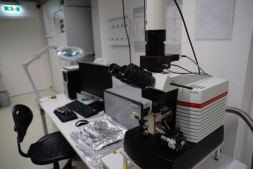
The recently installed VMQM is at the heart of the group's experimental activities. This unique instrument based on the Nion HERMES microscope by Bruker is an aberration-corrected and monochromated ultra-high vacuum scanning transmission electron microscope with operation voltages between 30 and 200 kV with a probe size of ca. 1 Ångström, allowing atomic-resolution microscopy and spectroscopy. Its objective area has been modified for experiments under controlled atmospheres between 1010 and 106 mbar and an high-vacuum transfer line directly connecting it to the CANVAS system.

Vienna Microscope for Quantum Materials (VMQM) ©Jani Kotakoski
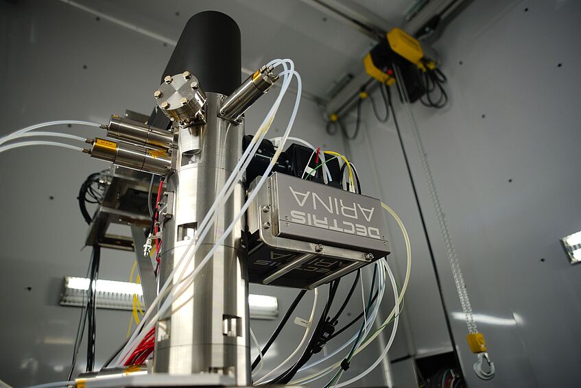
The Dectris ARINA is the latest-generation direct-electron camera that is optimized for ultrafast measurements at the lower primary beam energies. The Si-based sensor chip has 192×192 unusually large pixels of 100×100 μm in size, allowing it to accurately count every electron for any realistic electron-beam current. The speed of the camera is limited by the readout electronics' bandwidth of 10 Gb/s, resulting in a full-frame readout at 20 kHz or 2× binned and compressed readout at 120 kHz – corresponding to typical ADF scan rates. The ARINA thus enables uncompromising 4D-STEM measurements, bringing the group's experimental capabilities to the very leading edge of research worldwide.
For more information, see the Dectris website.
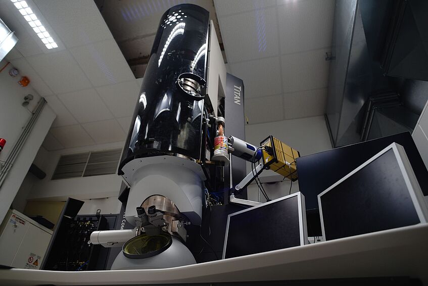
Transmission electron microscope from Thermo Fisher with an aberration corrector (CEOS Cs image corrector) an SFEG electron source, Gatan Tridiem 863 image filter for EELS, Oxford Instruments EDX system, and capabilities for tomography, scanning transmission electron microscopy, as well as liquid-phase and in-situ microscopy (mechanical, thermal and electric). Available acceleration voltages range from 80 to 300 kV (currently aligned at 80 kV, 200 kV, and 300 kV).
Delong Instruments Low-Voltage Electron Microscope (LVEM5) with an acceleration voltage of 5 kV. The microscope can be operated in transmission electron microscopy (TEM) and electron diffraction (ED) modes, as well as as an scanning electron microscope (SEM) and a scanning transmission electron microscope (STEM).